

MRI obtained to work up a sinonasal mass found in a patient w/ frequent epistaxis, what is the most likely diagnosis?
🔷More images in 🧵
#ENT #meded #medicine #Neurology #neurosurgery #FOAMed #radres #futreradres @AlbanyMedRadRes @ASHNRSociety
🔷More images in 🧵
#ENT #meded #medicine #Neurology #neurosurgery #FOAMed #radres #futreradres @AlbanyMedRadRes @ASHNRSociety
More images 👇
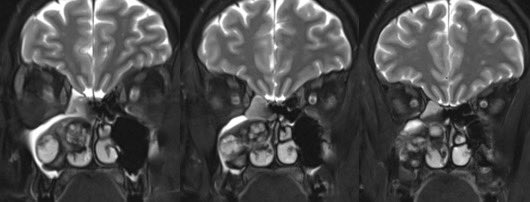


Answer: Sinonasal organizing hematoma
🔷These are uncommon hemorrhagic non-neoplastic masses that can be mistaken for malignancy
🔷CLINICAL: non specific epistaxis and nasal obstruction
🔷These are uncommon hemorrhagic non-neoplastic masses that can be mistaken for malignancy
🔷CLINICAL: non specific epistaxis and nasal obstruction
🔷Possible etiology (similar to cholesterol granuloma): Sinonasal hemorrhage into a closed cavity leads to negative pressure and repetitive rupture of friable vessels. Next is fibrosis, neovascularization w/ weak endothelium, and capsular formation which may prevent resorption
🔷ASSOCIATIONS: Coagulopathy, prior sinonasal surgery, ESRD, cirrhosis
🔷LOCATION: Maxillary sinus most common (usually medial near ostium)
🔷LOCATION: Maxillary sinus most common (usually medial near ostium)
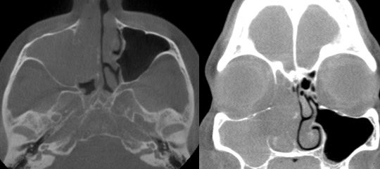
🔷IMAGING:
CT: relatively non specific w/ expansile heterogeneously dense sinonasal mass often w/ smooth bony erosions
CT: relatively non specific w/ expansile heterogeneously dense sinonasal mass often w/ smooth bony erosions

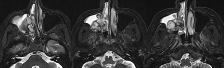
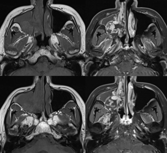
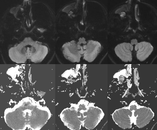
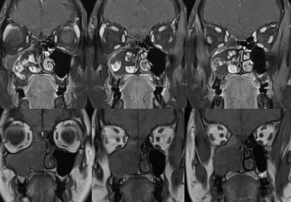
MRI:
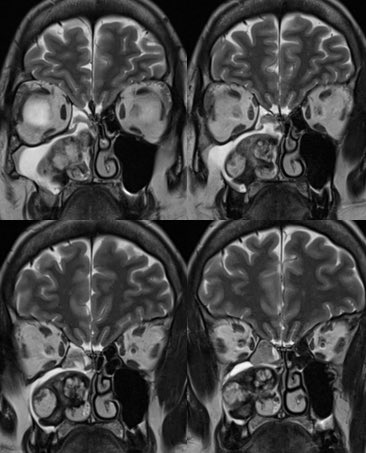
▶️WELL DEMARCATED from surrounding structures
▶️Marked HETEROGENEOUS signal on T2 surrounded by PERIPHERAL T2 DARK RIM
▶️Marked irregular papillary or frond-like ENHANCEMENT from the neovascularity
▶️T1 also heterogenous w/ some areas high signal
▶️WELL DEMARCATED from surrounding structures
▶️Marked HETEROGENEOUS signal on T2 surrounded by PERIPHERAL T2 DARK RIM
▶️Marked irregular papillary or frond-like ENHANCEMENT from the neovascularity
▶️T1 also heterogenous w/ some areas high signal


pubs.rsna.org/doi/10.1148/ra…
Case 217: Sinonasal Organized Hematoma | Radiology
History A 15-year-old boy presented to the emergency department with intractable epistaxis. He had a...

ncbi.nlm.nih.gov/pmc/articles/P…
Sinonasal Organized Hematoma: CT and MR Imaging Findings
BACKGROUND AND PURPOSE: Sinonasal organized hematoma (OH) is an uncommon, nonneoplastic benign condi...

ajronline.org/doi/full/10.22…
Organized Hematoma of the Maxillary Sinus: CT Findings | AJR
OBJECTIVE. Organized hematoma of the maxillary sinus is rare. It occurs by organization of the hemat...
ajnr.org/ajnr-case-coll…
Sinonasal Organized Hematoma | American Journal of Neuroradiology
Loading suggestions...