

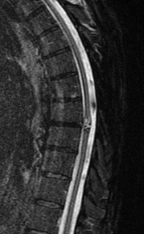
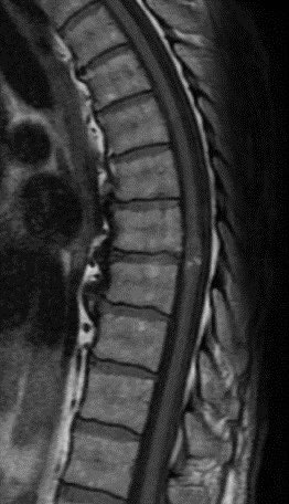
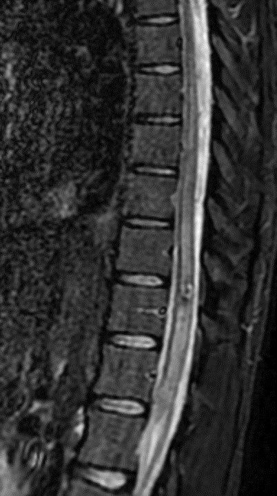
What is the most likely diagnosis in this 65 y/o M presenting w/ leg weakness, hyperreflexia, spasticity and absent vibration sense in the LE?
#neurosurgery #neurology #meded #medicine #futureradres #radres @The_ASSR
#neurosurgery #neurology #meded #medicine #futureradres #radres @The_ASSR
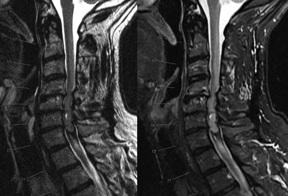
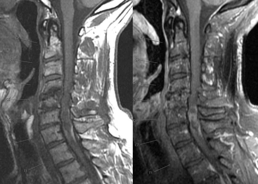


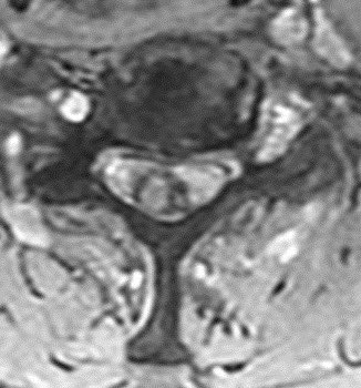
Answer: Spinal ependymoma grade 2 showing the hemosiderin cap sign (the dark signal on T2, STIR and GRE at the superior and inferior edges of the tumor)
💡 Ependymomas and astrocytomas constitute ~70% of intramedullary spinal cord tumors
💡 Ependymomas and astrocytomas constitute ~70% of intramedullary spinal cord tumors
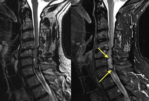
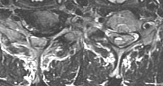
🔷Typical Spinal Ependymomas:
▶️More common in ADULTS & NF2
▶️More WELL DEFINED margins
▶️CENTRAL location expanding outward (since they arise from the ependymal lining along the central canal of the spinal cord)
▶️Polar cysts and HEMOSIDERIN CAP SIGN
▶️More common in ADULTS & NF2
▶️More WELL DEFINED margins
▶️CENTRAL location expanding outward (since they arise from the ependymal lining along the central canal of the spinal cord)
▶️Polar cysts and HEMOSIDERIN CAP SIGN


🔷Typical Spinal Astrocytomas:
▶️More common in CHILDREN & NF1
▶️INFILTRATING since they arise in the parenchyma (though can be circumscribed usually when low grade)
▶️ECCENTRIC rather than central in spinal cord (though tough to tell considering the cord is only ~1cm thick)
▶️More common in CHILDREN & NF1
▶️INFILTRATING since they arise in the parenchyma (though can be circumscribed usually when low grade)
▶️ECCENTRIC rather than central in spinal cord (though tough to tell considering the cord is only ~1cm thick)
💡 Maximal safe resection mostly depends on preexisting neuro deficits and intraoperative dissection planes. However, assessing likelihood of a circumscribed vs infiltrating tumor can help strategize resection or biopsy
💡It is also important to try and differentiate tumoral cysts (which usually peripherally enhance) vs non tumoral cysts (which do not peripherally enhance)
💡 Tumoral cysts should be removed or the tumor may recur while non tumoral cysts can be decompressed or left behind. This is especially important while attempting maximal safe resection in such a small space
🔷Less common intramedullary tumors include:
▶️hemangioblastoma (arise at pial surface, flow voids, cyst with enhancing nodule, can be sporadic though ⬆️ incidence in VHL)
▶️intramedullary myxopapillary ependymoma (usually intradural extramedullary but can manifest in the conus)
▶️hemangioblastoma (arise at pial surface, flow voids, cyst with enhancing nodule, can be sporadic though ⬆️ incidence in VHL)
▶️intramedullary myxopapillary ependymoma (usually intradural extramedullary but can manifest in the conus)
🔷Less common intramedullary tumors cont’d:
▶️Mets (rare usually lung, breast or melanoma in setting of widespread metastatic disease)
▶️subependymoma (very rare, usually don’t enhance, have a subpial or eccentric growth pattern producing the bamboo leaf sign)
▶️Mets (rare usually lung, breast or melanoma in setting of widespread metastatic disease)
▶️subependymoma (very rare, usually don’t enhance, have a subpial or eccentric growth pattern producing the bamboo leaf sign)
🔷Other differentials include Cavernoma, epidermoid cyst and inflammatory or infectious myelitis. It is important to take a step back and ask yourself if you could be dealing with an inflammatory/infectious myelitis
💡 Consider short follow up to eval for change and CSF analysis
💡 Consider short follow up to eval for change and CSF analysis
⭐️ Two companion cases of a spinal Cavernoma (one with cord edema in last image)
▶️Cavernomas have a similar popcorn appearance as seen in the brain and should not have solid or nodular enhancing components though mild enhancement can be seen
▶️Cavernomas have a similar popcorn appearance as seen in the brain and should not have solid or nodular enhancing components though mild enhancement can be seen


Loading suggestions...