

2/Specific patterns of atrophy are associated w/Alzheimer’s disease (AD).
Atrophy often involves the hippocampus & the anterior/medial portion of the adjacent parahippocampal gyrus, called the entorhinal cortex
This is where to should look for volume loss on imaging, but how?
Atrophy often involves the hippocampus & the anterior/medial portion of the adjacent parahippocampal gyrus, called the entorhinal cortex
This is where to should look for volume loss on imaging, but how?

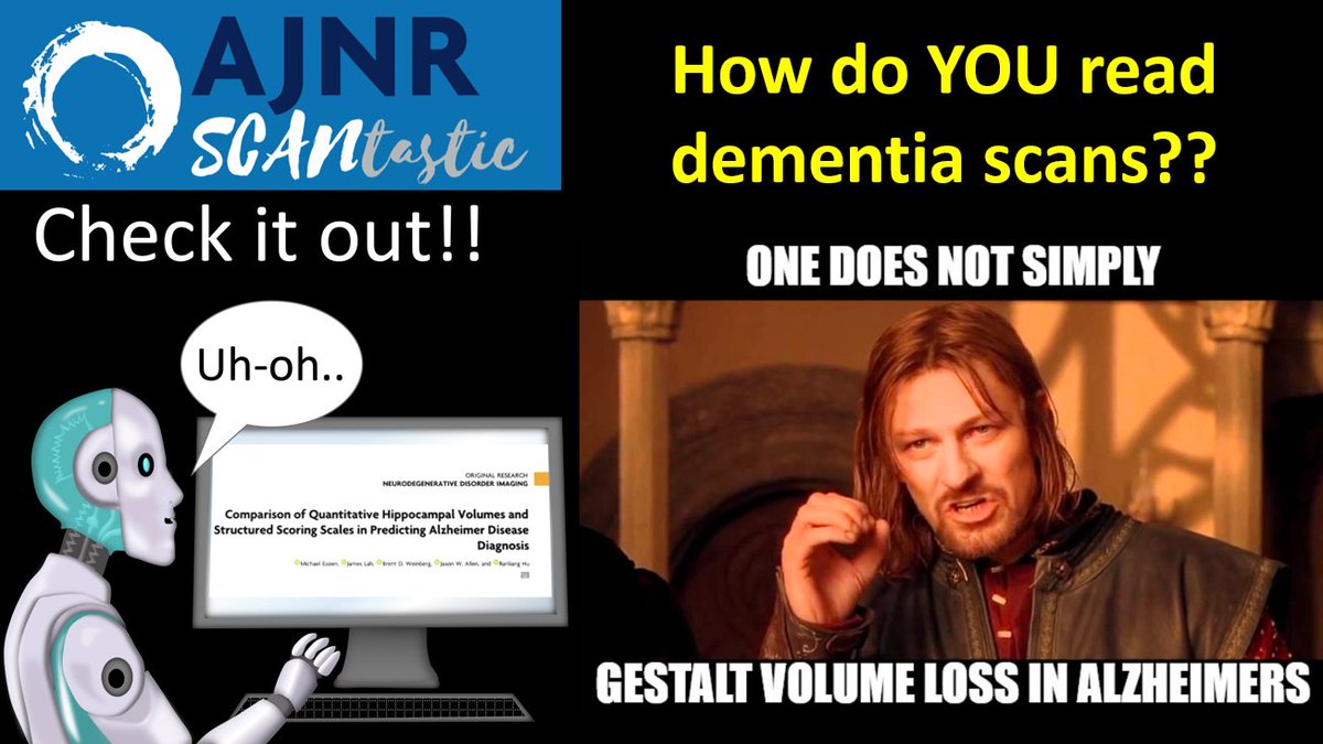
3/Many people do just gestalt the atrophy. But this isn't reproducible.
Others rely on automated software to give quantitative volumes down to the 100th of a mL & compare them w/an age-matched database.
Many feel this is superior—because who WOULDN’T want more data?
Others rely on automated software to give quantitative volumes down to the 100th of a mL & compare them w/an age-matched database.
Many feel this is superior—because who WOULDN’T want more data?
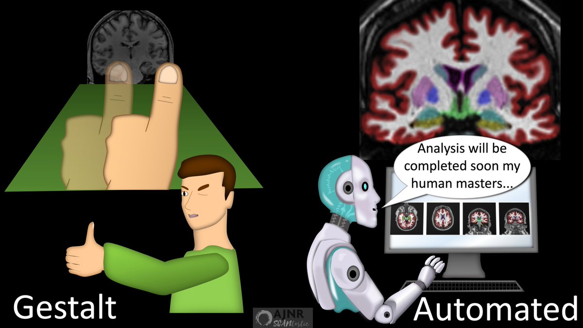
4/However, automated machine analysis is not always better.
There can be segmentation errors affect the analysis.
And even small segmentation errors could have a significant impact when the volumes in this region are already so small
There can be segmentation errors affect the analysis.
And even small segmentation errors could have a significant impact when the volumes in this region are already so small

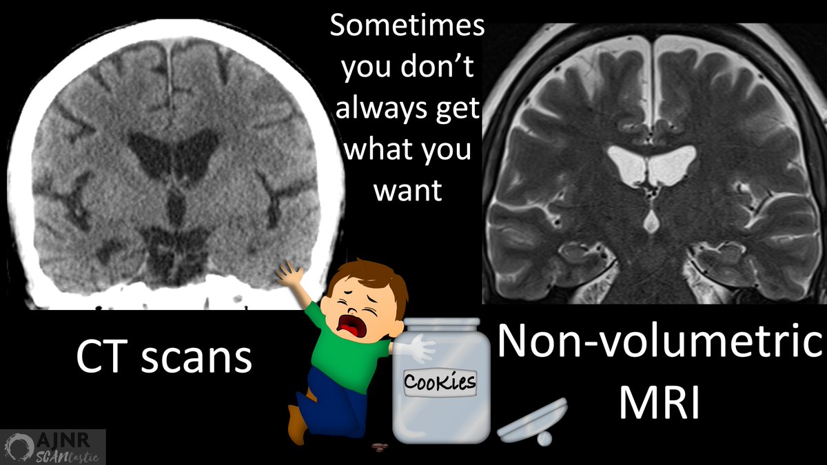
5/Also, automated analysis can only be used on studies with volumetric imaging
But often times you have studies like CTs or routine MRIs that you would like to evaluate for possible AD.
Here, automated analysis cannot help you.
But often times you have studies like CTs or routine MRIs that you would like to evaluate for possible AD.
Here, automated analysis cannot help you.
6/But gestalt-ing it isn’t the answer either--can be inaccurate & isn't reproducible.
Maybe the answer is somewhere in between.
2 very reproducible semiquantitative scores exist: medial temporal atrophy score (MTA) & entorhinal cortex atrophy score (ERICA)
How do we use them?
Maybe the answer is somewhere in between.
2 very reproducible semiquantitative scores exist: medial temporal atrophy score (MTA) & entorhinal cortex atrophy score (ERICA)
How do we use them?

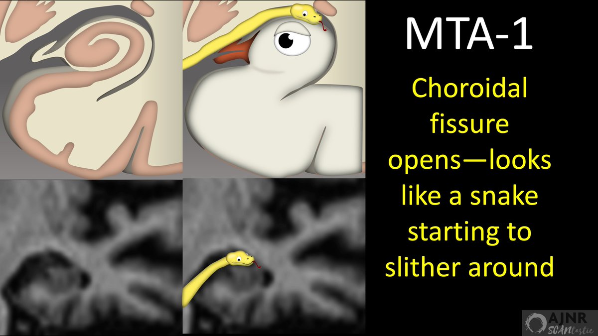
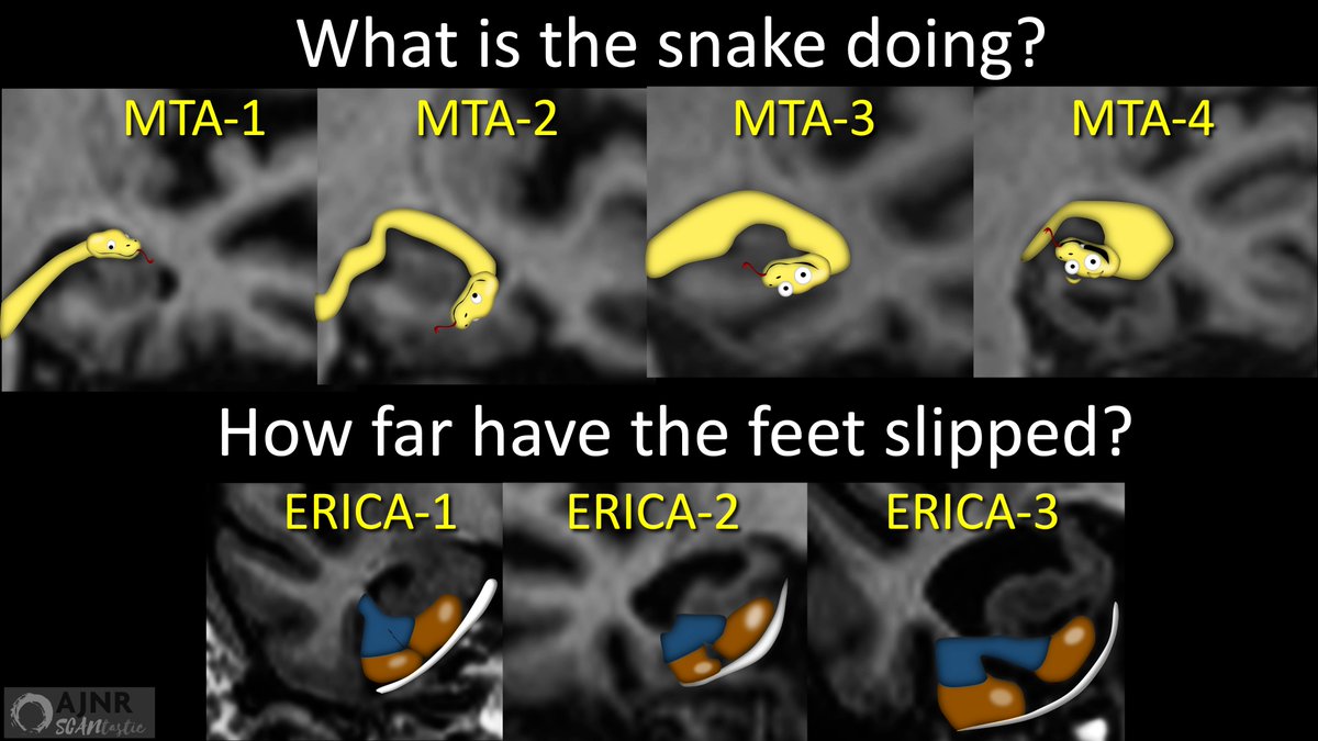
7/Let’s start w/the MTA score.
On coronal images, the hippocampus looks like a bird.
Typically, there is very little room around the bird’s head.
But w/volume loss, CSF slithers around like a snake.
MTA score grades how far the snake has gone towards eating the bird!
On coronal images, the hippocampus looks like a bird.
Typically, there is very little room around the bird’s head.
But w/volume loss, CSF slithers around like a snake.
MTA score grades how far the snake has gone towards eating the bird!

8/MTA-1 is the first step a snake takes towards eating the bird.
Here the choroidal fissure has slightly widened.
So, as a first step, the snake has begun to slither around the head of the bird.
Here the choroidal fissure has slightly widened.
So, as a first step, the snake has begun to slither around the head of the bird.
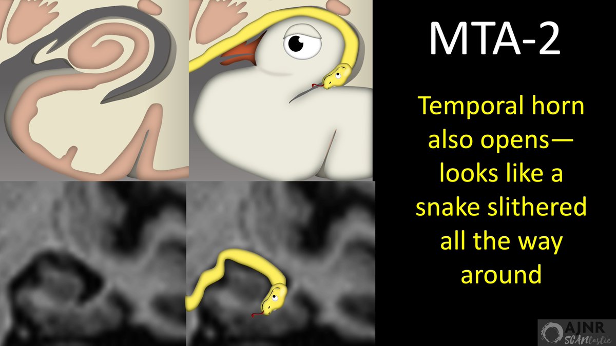
9/For MTA-2, the temporal horn also widens & CSF can be seen encircling the entire top of the hippocampus.
Now the snake has taken the next step to encircle the head of it’s bird victim.
Now the snake has taken the next step to encircle the head of it’s bird victim.
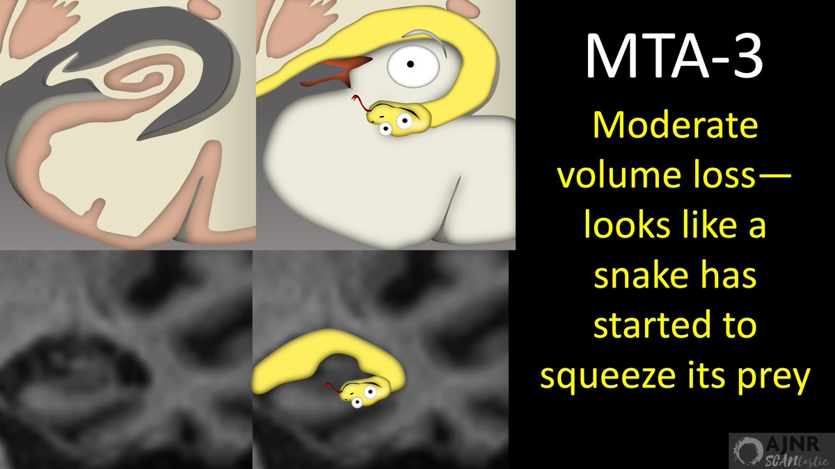
10/In MTA-3, the hippocampus now shows moderate volume loss—so it looks like the head of the bird has been compressed.
So now it looks like the snake has not only encircled the head of the bird, but it has begun the final steps to squeeze down as well.
So now it looks like the snake has not only encircled the head of the bird, but it has begun the final steps to squeeze down as well.
11/For MTA-4, the hippocampal volume loss is profound, so it looks like the head of bird has been crushed by the snake.
So now, the snake has completed its attack, with each step corresponding to an MTA score:
(1) Slithering in
(2) Encircling
(3) Squeezing
(4) Crushing
So now, the snake has completed its attack, with each step corresponding to an MTA score:
(1) Slithering in
(2) Encircling
(3) Squeezing
(4) Crushing

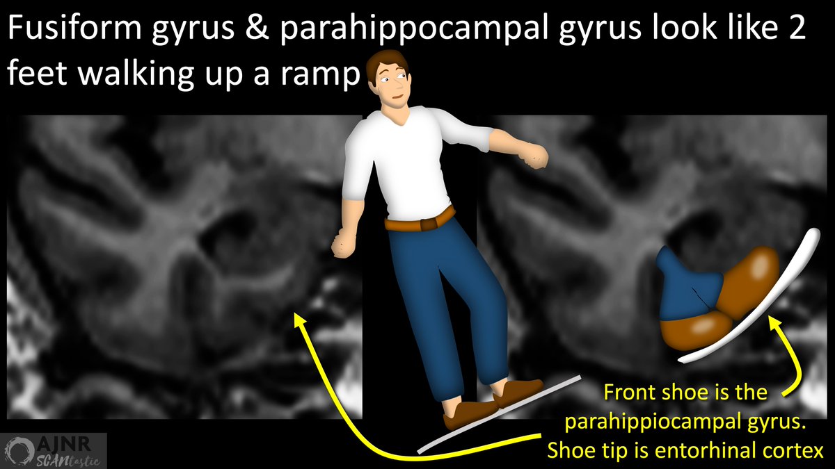
12/Now for the entorhinal cortex atrophy or ERICA score.
It is evaluated at the level of the mamillary bodies.
At this level, the parahippocampal gyrus & the adjacent fusiform gyrus look like two shows going up a ramp.
Entorhinal cortex is the tip of the leading shoe.
It is evaluated at the level of the mamillary bodies.
At this level, the parahippocampal gyrus & the adjacent fusiform gyrus look like two shows going up a ramp.
Entorhinal cortex is the tip of the leading shoe.
13/Entorhinal cortex atrophy looks like the feet have slipped on a banana
In the most mild case, your front foot slips a little separating your legs
Same w/the entorhinal cortex.
Volume loss widens the collateral sulcus here, so it looks like the legs slipped apart a bit
In the most mild case, your front foot slips a little separating your legs
Same w/the entorhinal cortex.
Volume loss widens the collateral sulcus here, so it looks like the legs slipped apart a bit

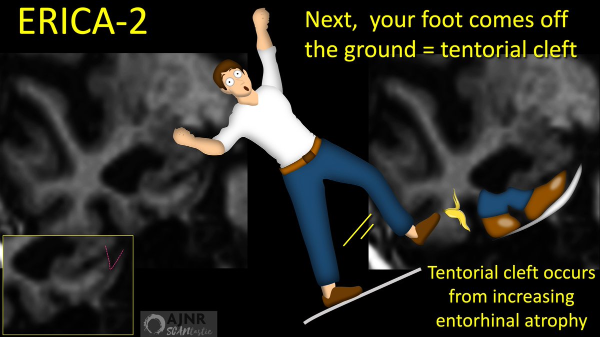
14/If your slip was a little worse, your foot is going to fly into the air.
Same w/the entorhinal cortex.
Now, the entorhinal cortex loses enough volume so there is now CSF between it & the tentorium (tentorial cleft sign)
It looks like a uplifted foot during a slip!
Same w/the entorhinal cortex.
Now, the entorhinal cortex loses enough volume so there is now CSF between it & the tentorium (tentorial cleft sign)
It looks like a uplifted foot during a slip!
15/Finally, with a really bad slip, you are doing the full splits in the air.
It’s same look for bad entorhinal cortex volume loss.
There’s so much volume loss that the collateral sulcus doesn’t look like a small gap between the legs—it looks like a full on splits!
It’s same look for bad entorhinal cortex volume loss.
There’s so much volume loss that the collateral sulcus doesn’t look like a small gap between the legs—it looks like a full on splits!

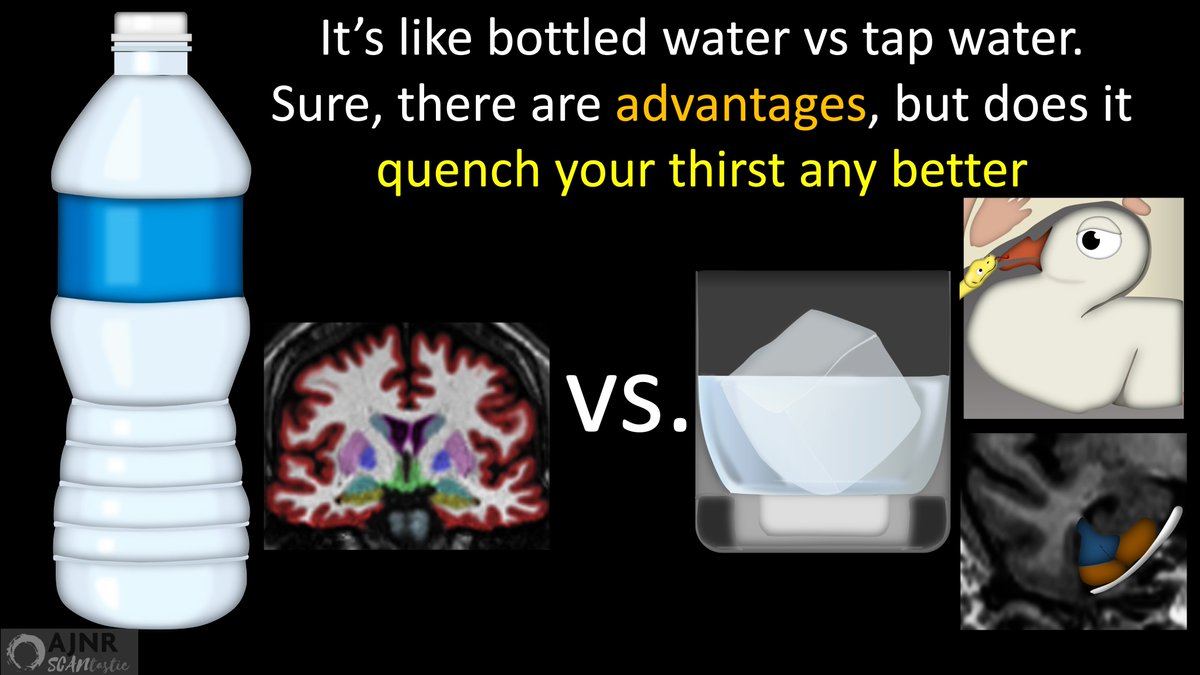
16/In this month’s @theAJNR, Essien et al. compared these semiquantitative scores w/the automated software analysis.
They found that while there were small differences, overall, quantitative volumes & qualitative scores performed very similarly diagnostically.
They found that while there were small differences, overall, quantitative volumes & qualitative scores performed very similarly diagnostically.

17/Automated analysis is like bottled water
It’s more expensive, but has advantages, like convenient packaging. But for what matters, it doesn’t quench thirst better than tap water
Automated analysis is convenient & quantitative. But for diagnosis, it doesn’t do more than you
It’s more expensive, but has advantages, like convenient packaging. But for what matters, it doesn’t quench thirst better than tap water
Automated analysis is convenient & quantitative. But for diagnosis, it doesn’t do more than you
18/So for every study you read for dementia or memory loss or even for falls, never FORGET to yourself two questions:
(1) How far is the snake into his attack on the bird?
(2) How far have the shoes slipped on a banana?
That’s the analysis you need!
(1) How far is the snake into his attack on the bird?
(2) How far have the shoes slipped on a banana?
That’s the analysis you need!
جاري تحميل الاقتراحات...