

1/Sometimes the tiniest thing can be the biggest pain—that’s microvascular compression of the trigeminal nerve! But seeing such a tiny finding can be hard!
Here’s a #tweetorial about how to look at the trigeminal nerve on MRI
#medtwitter #neurotwitter #neurorad #meded #FOAMed
Here’s a #tweetorial about how to look at the trigeminal nerve on MRI
#medtwitter #neurotwitter #neurorad #meded #FOAMed
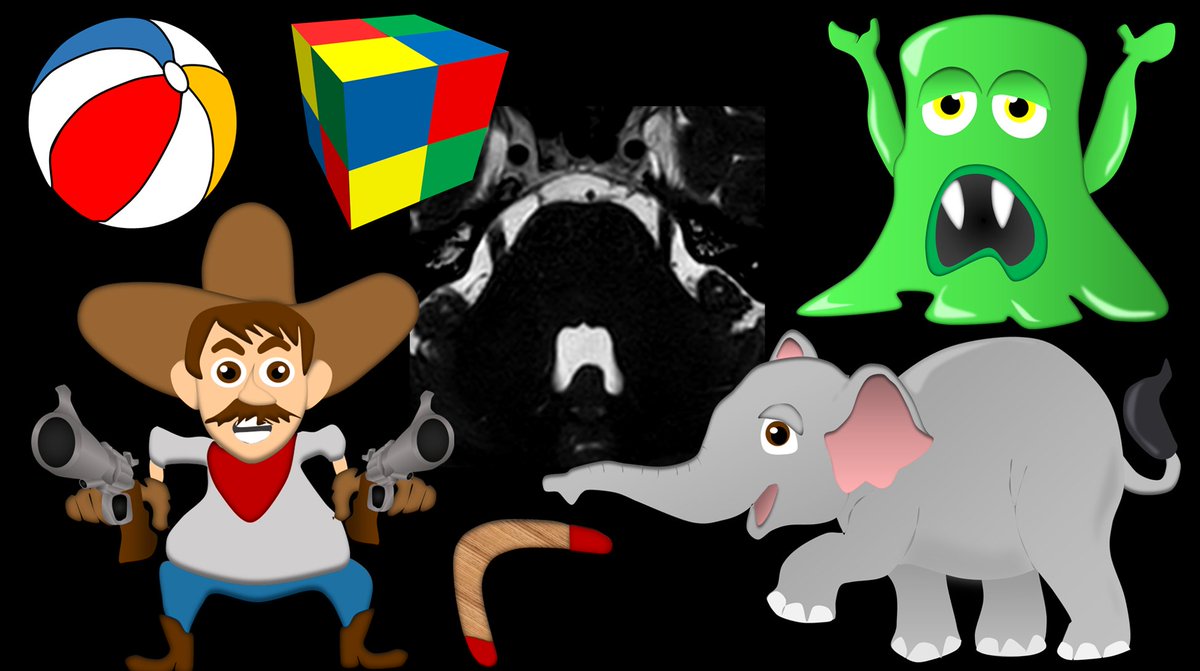
2/The most important thing to remember is that the nerve is 3D so you have to look for compression in all 3 planes. Let’s start w/the axial plane. On a normal axial, the trigeminal nerves should look like the arms of an alien sticking out of the pons.
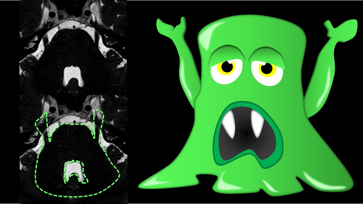
3/Compression in the axial plane usually will deviate the nerve laterally—so that the Alien looks like he is flexing one of his arms. So if you see the Alien trying to show his guns—that’s microvascular compression!
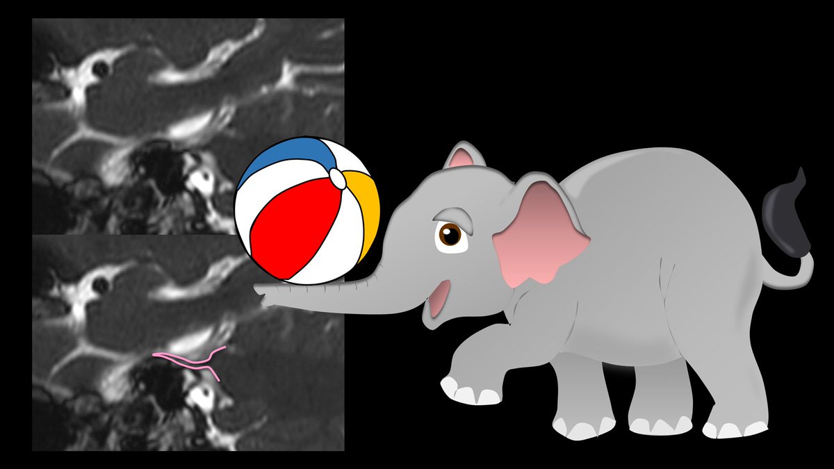
4/In the sagittal plane, the nerve looks like an elephant’s trunk coming out of the pons. It should have a smooth curve up and over before it enters Meckel’s cave, just like the way an elephant’s trunk curves.
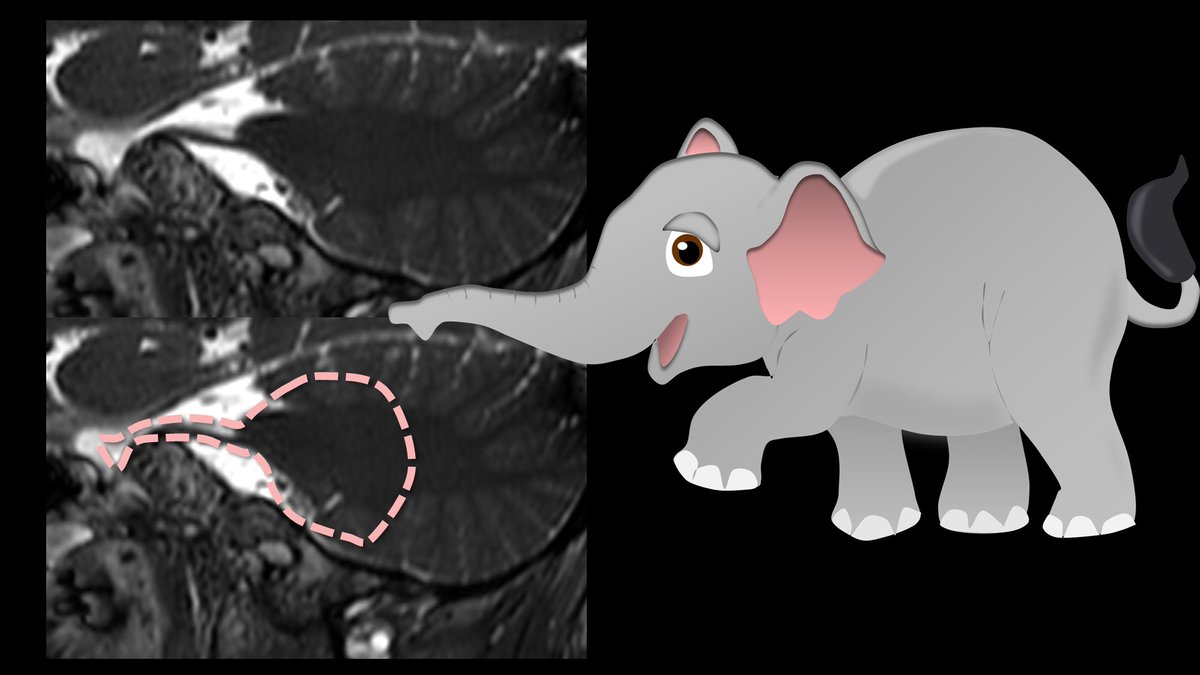
5/If the trunk is flattened, like it’s balancing ball or is curved downwards—that’s microvascular compression in the sagittal plane. If the nerve is pressed down, you could miss this on the axial plane—bc the movement is parallel to the axial plane. You need a sagittal view.
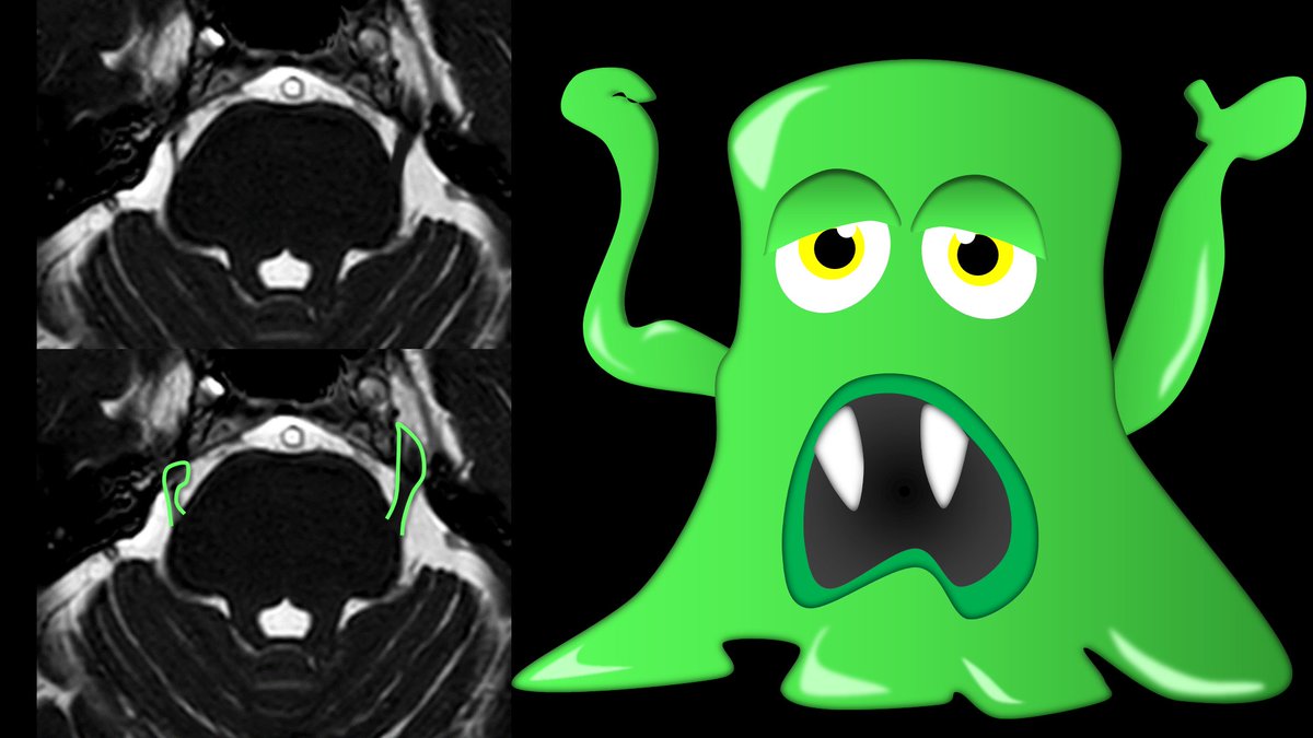
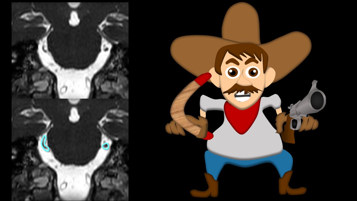
6/In the coronal plane, the nerves look like two gun barrels pointed at you, by a very potty bellied cowboy that is the pons.
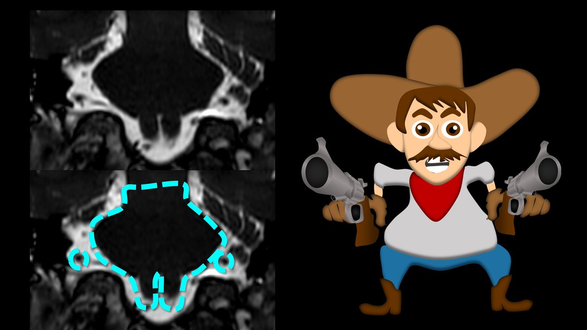
7/If the nerves lose their gun barrel shape, and look more like a boomerang—in any direction—that is microvascular compression. Coronal is usually the most helpful view, bc you can see movement both up and down and left to right.
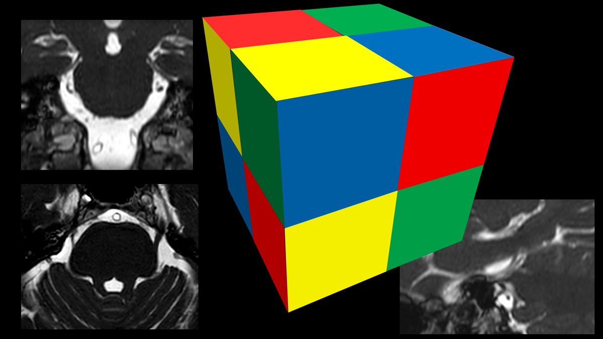
8/So now you know what the normal trigeminal nerve looks like in all 3 planes—and now you can check for microvascular compression in three dimensions. Remember, images may be 2D, but life—and pathology—are 3D!
Loading suggestions...