

💀 Pulmonary Infarction 💀
Is that focal chest consolidation a pneumonia? 🤔
Let's talk about a pneumonia mimicker that can be *very* easily missed if you've never seen it before 🎭
Is that focal chest consolidation a pneumonia? 🤔
Let's talk about a pneumonia mimicker that can be *very* easily missed if you've never seen it before 🎭

Pulmonary Infarction is the result of a simple series of events:
🩸 Pulmonary artery obstruction
↓
🩸 Alveolar hemorrhage & edema
☠️ If fluid not absorbed, RBC lysis & hemosiderin release leads to lung necrosis! ☠️
🩸 Pulmonary artery obstruction
↓
🩸 Alveolar hemorrhage & edema
☠️ If fluid not absorbed, RBC lysis & hemosiderin release leads to lung necrosis! ☠️
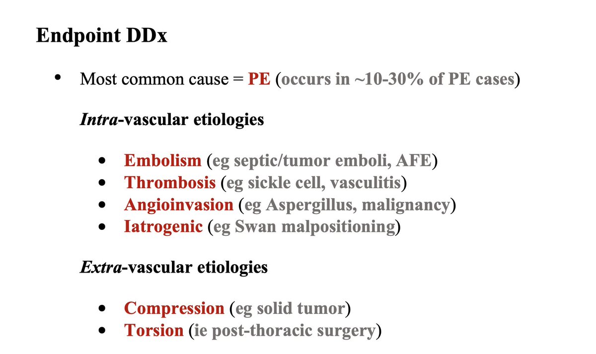
Given this pathophysiology, Pulmonary Infarction can be due to any etiology resulting in PA arterial obstruction. The most common cause is PE (occurring in ~10-30% of PE cases)!
For other etiologies, break down into intra- vs. extra-vascular etiologies to find the Endpoint Dx:
For other etiologies, break down into intra- vs. extra-vascular etiologies to find the Endpoint Dx:
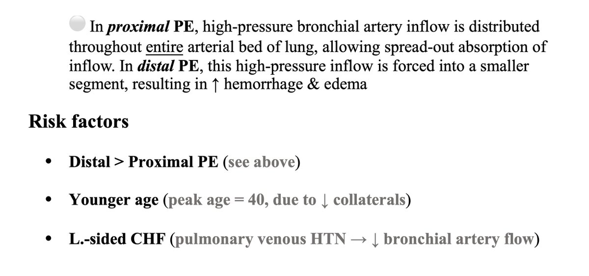
Risk factors include:
🔥 Distal PEs (because high-pressure bronchial artery inflow is forced into a smaller segment of lung → ↑ hemorrhage & edema)
🔥 Younger age (due to ↓ collaterals)
🔥 L.-sided CHF (pulmonary venous HTN → ↓ bronchial artery flow)
🔥 Distal PEs (because high-pressure bronchial artery inflow is forced into a smaller segment of lung → ↑ hemorrhage & edema)
🔥 Younger age (due to ↓ collaterals)
🔥 L.-sided CHF (pulmonary venous HTN → ↓ bronchial artery flow)
Symptoms can be quite variable! Think parenchymal & pleural irritation leading to:
- Dyspnea (~70-80%) & Cough (Pleuritic symptoms)
- Chest pain (~50-70%) & Back pain (Due to anterior vs. posterior lung infarction)
- Hemoptysis (~5-20%)
- Fever (~5-10%)
- Dyspnea (~70-80%) & Cough (Pleuritic symptoms)
- Chest pain (~50-70%) & Back pain (Due to anterior vs. posterior lung infarction)
- Hemoptysis (~5-20%)
- Fever (~5-10%)
OK, so those symptoms are somewhat non-specific 🤔
Chest imaging findings serve as invaluable Dx pivots.
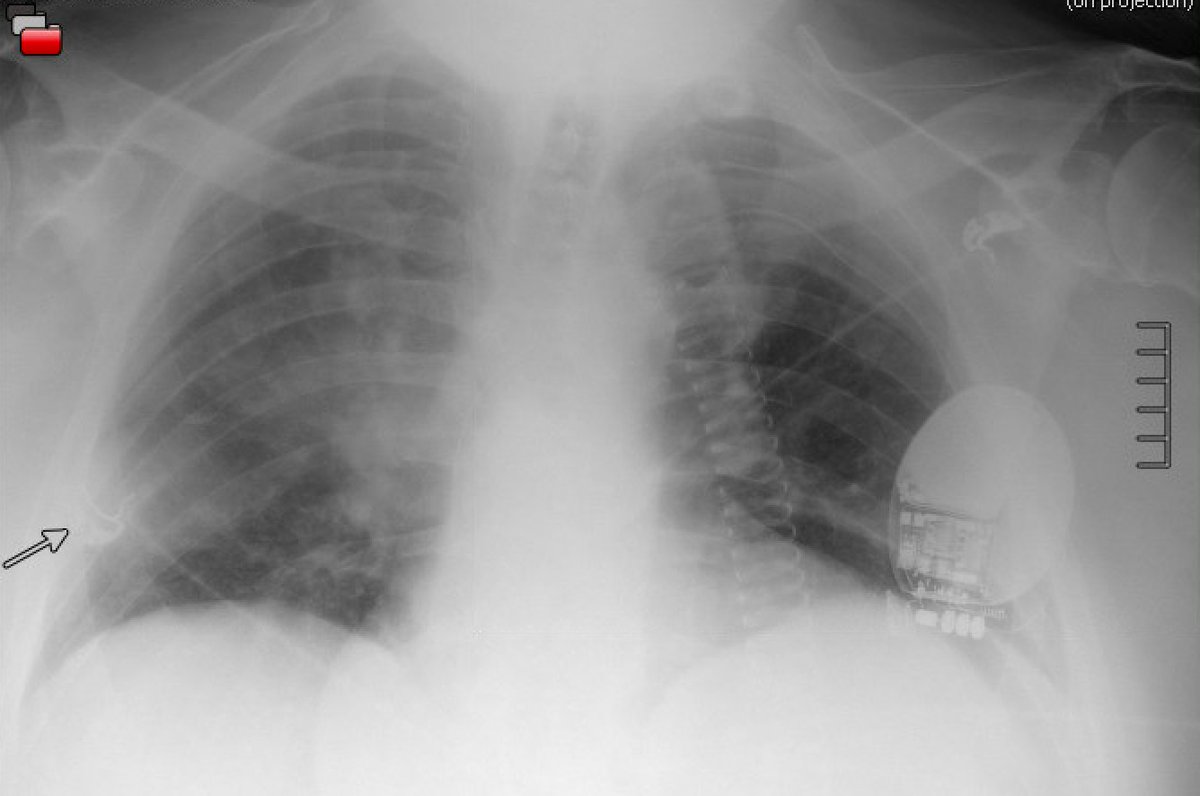
CXR findings:
☠️ Wedge-shaped opacity ("Hampton's hump")
☠️ ↑ Peripheral lucency ("Westermark's sign")
☠️ ↑ PA size ("Fleischner sign")
Chest imaging findings serve as invaluable Dx pivots.
CXR findings:
☠️ Wedge-shaped opacity ("Hampton's hump")
☠️ ↑ Peripheral lucency ("Westermark's sign")
☠️ ↑ PA size ("Fleischner sign")
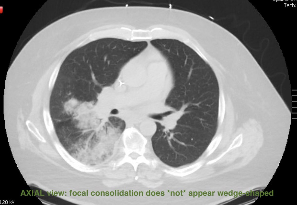
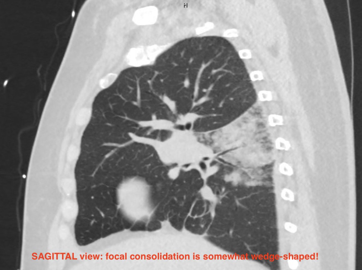
CT findings:
☠️ Wedge vs. Semicircular-shaped focal consolidation
☠️ Internal air lucencies → Cavitation ("bubbly" consolidation)
☠️ Feeding vessel
☠️ Wedge vs. Semicircular-shaped focal consolidation
☠️ Internal air lucencies → Cavitation ("bubbly" consolidation)
☠️ Feeding vessel
📢 Do your own review of CT imaging in all views, or you can miss these findings! These findings may be read as "pneumonia" by radiology! The wedge shape may not be obvious in one view, but may be obvious in another view! 📢
Here's my proposed Dx approach:
- (+) Focal CXR or CT chest finding?
↓
- Clinical picture = Pneumonia?
✅ Yes → Pneumonia prioritized
❌ No → CT PE rule-out
- (+) Focal CXR or CT chest finding?
↓
- Clinical picture = Pneumonia?
✅ Yes → Pneumonia prioritized
❌ No → CT PE rule-out
🏁 Final pearls:
⚪ A discordant CRP (high) & Procalcitonin (low) in the presence of a suspicious focal chest CT finding & history *inconsistent* with pneumonia may serve as a Dx pivot
⚪ Don't forget, fever can be due to thrombosis!
⚪ A discordant CRP (high) & Procalcitonin (low) in the presence of a suspicious focal chest CT finding & history *inconsistent* with pneumonia may serve as a Dx pivot
⚪ Don't forget, fever can be due to thrombosis!
⚪ Stay curious, my friends: this Dx is extraordinarily hard to make if it's not on your radar. This illness script was born out of a case of infarction that went misdiagnosed for >1 week due to radiology read of ⚓ "pneumonia" ⚓ . Of note, presenting sxs may be *chronic*!
🏁 Hopefully you enjoyed & please amplify this challenging Dx!
CC: @Sharminzi @rabihmgeha @DxRxEdu @thilanMD @EM_RESUS @marywhite_md @medrants @nsrosenberg @kiaracamacho96 @minheredia @ArcieriMichael @TLHM_MD @MDVictorJimenez @Heard_that_alex @MarkDSiegel1 @MadellenaC
CC: @Sharminzi @rabihmgeha @DxRxEdu @thilanMD @EM_RESUS @marywhite_md @medrants @nsrosenberg @kiaracamacho96 @minheredia @ArcieriMichael @TLHM_MD @MDVictorJimenez @Heard_that_alex @MarkDSiegel1 @MadellenaC
Save this Illness Script here: glass.health
جاري تحميل الاقتراحات...