

1/ Time for another #EmoryNCCTweetorials!
The setup: Grady Hospital at @EmoryNeuroCrit
The pt: Jane Doe coming through the ED as a stroke alert
Another stroke you say?… @MedTweetorials
#MedEd #tweetorial #neurotwitter
The setup: Grady Hospital at @EmoryNeuroCrit
The pt: Jane Doe coming through the ED as a stroke alert
Another stroke you say?… @MedTweetorials
#MedEd #tweetorial #neurotwitter

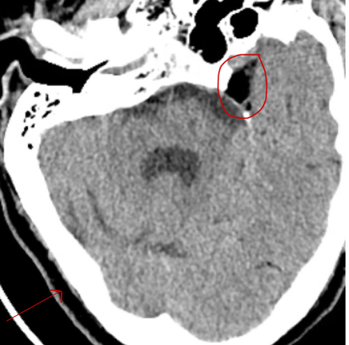
2/ She presents with L face/arm/leg weakness with her CT scan shown below
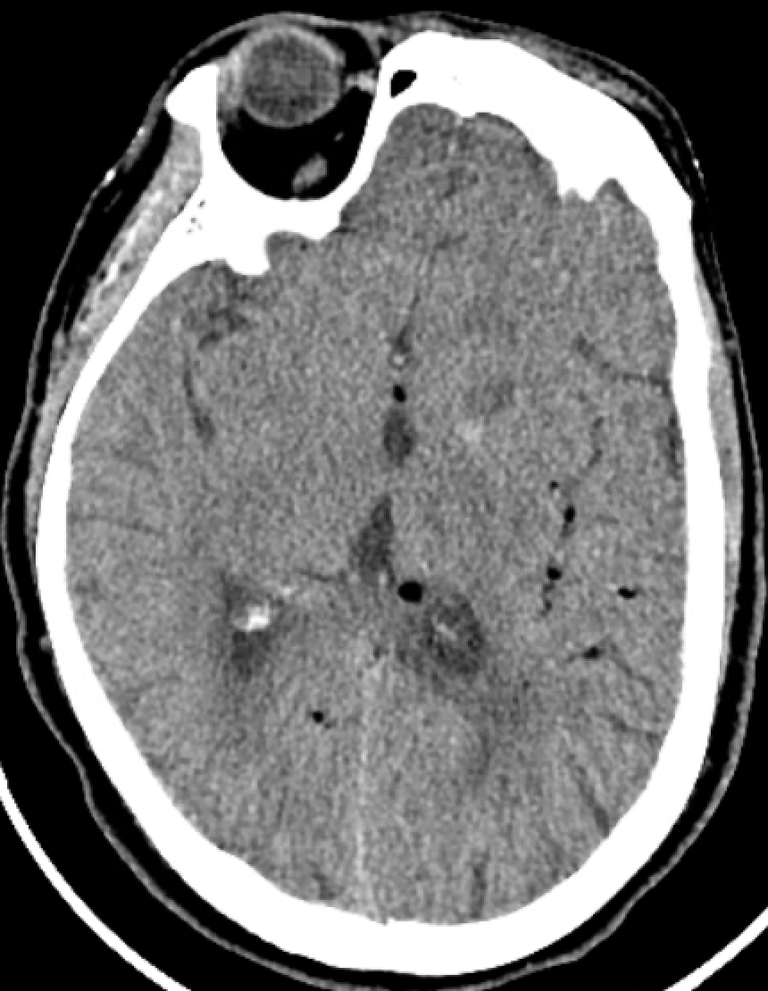
3/ Doesn't look too bad.
Wait a second…why are there black spots (orange arrow) everywhere?
Wait a second…why are there black spots (orange arrow) everywhere?
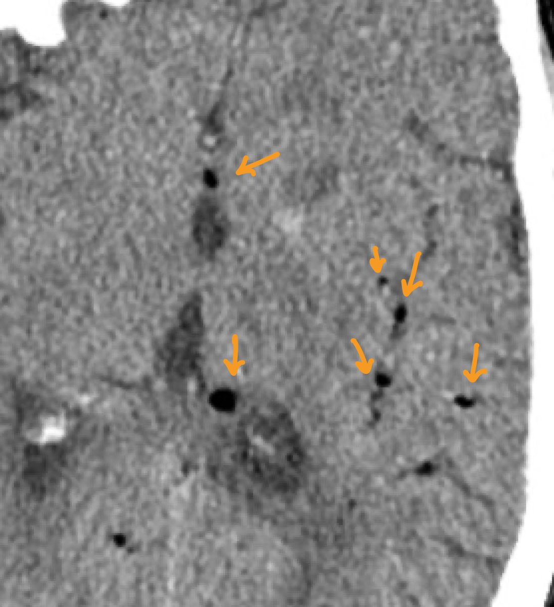
4/ Uh oh. So what are those hypodensities 🤔?
5/ Definitely an artifact 😅...maybe if we ignore it, the radiologists will ignore it on the final read as well (too bad they 👀 everything 🤦♂️)

6/ You decide to go back and check on the patient to double check that nothing was missed.
✅ Trauma?...Nope
✅ Recent surgeries near the head/neck?...Nope
✅ Central line or peripheral IV that might have gone arterial?...Nope
✅ Trauma?...Nope
✅ Recent surgeries near the head/neck?...Nope
✅ Central line or peripheral IV that might have gone arterial?...Nope

7/ Okay I know what it is for sure.
✅ CSF leak from nostrils/sinuses!
.
.
.
Nope, no leak 😭
✅ CSF leak from nostrils/sinuses!
.
.
.
Nope, no leak 😭

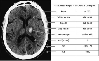
8/ You slowly remember being able to differentiate the density of materials using Hounsfield units (HU). But what the heck does a HU even mean?

9/ Hounsfield is a dimensionless unit and named after the inventor of the CT, Sir Godfrey Hounsfield.
It reflects the electron density at a given location and converted into a digital image by assigning a gray-scale intensity to each value
It reflects the electron density at a given location and converted into a digital image by assigning a gray-scale intensity to each value

10/ The higher the number, the brighter the pixel intensity.
Water is set at 0 HU with denser material being more positive (and thus lighter), whereas less dense material is more negative (and thus darker).
Water is set at 0 HU with denser material being more positive (and thus lighter), whereas less dense material is more negative (and thus darker).
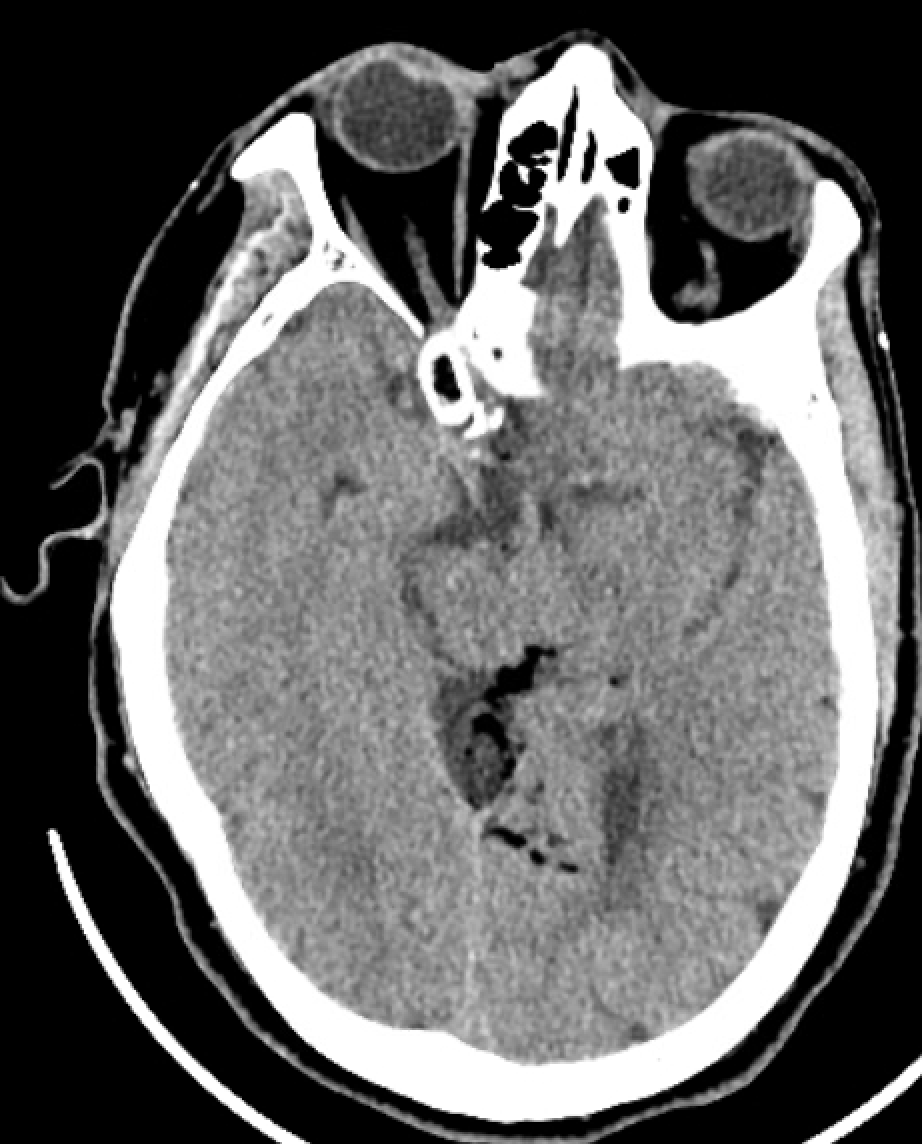
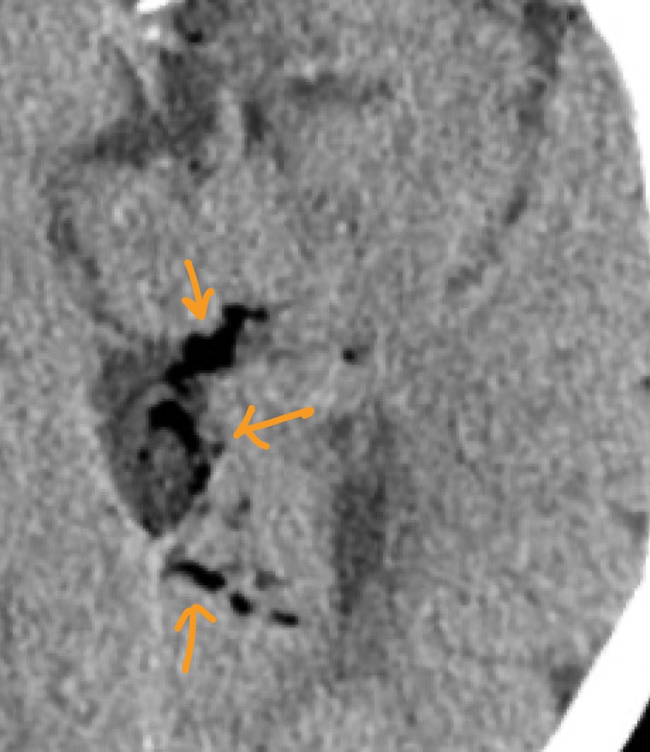
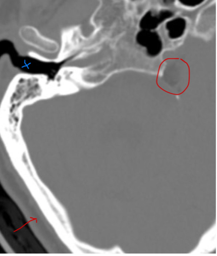
11/ Using this knowledge, we 👀 the area of interest (red circle) has the same density as subcutaneous fat (red arrow). Once we window the scan appropriately, we begin to see the different density between fat and air (blue x).
12/ But how did it get there? Her exam has been unrevealing thus far.

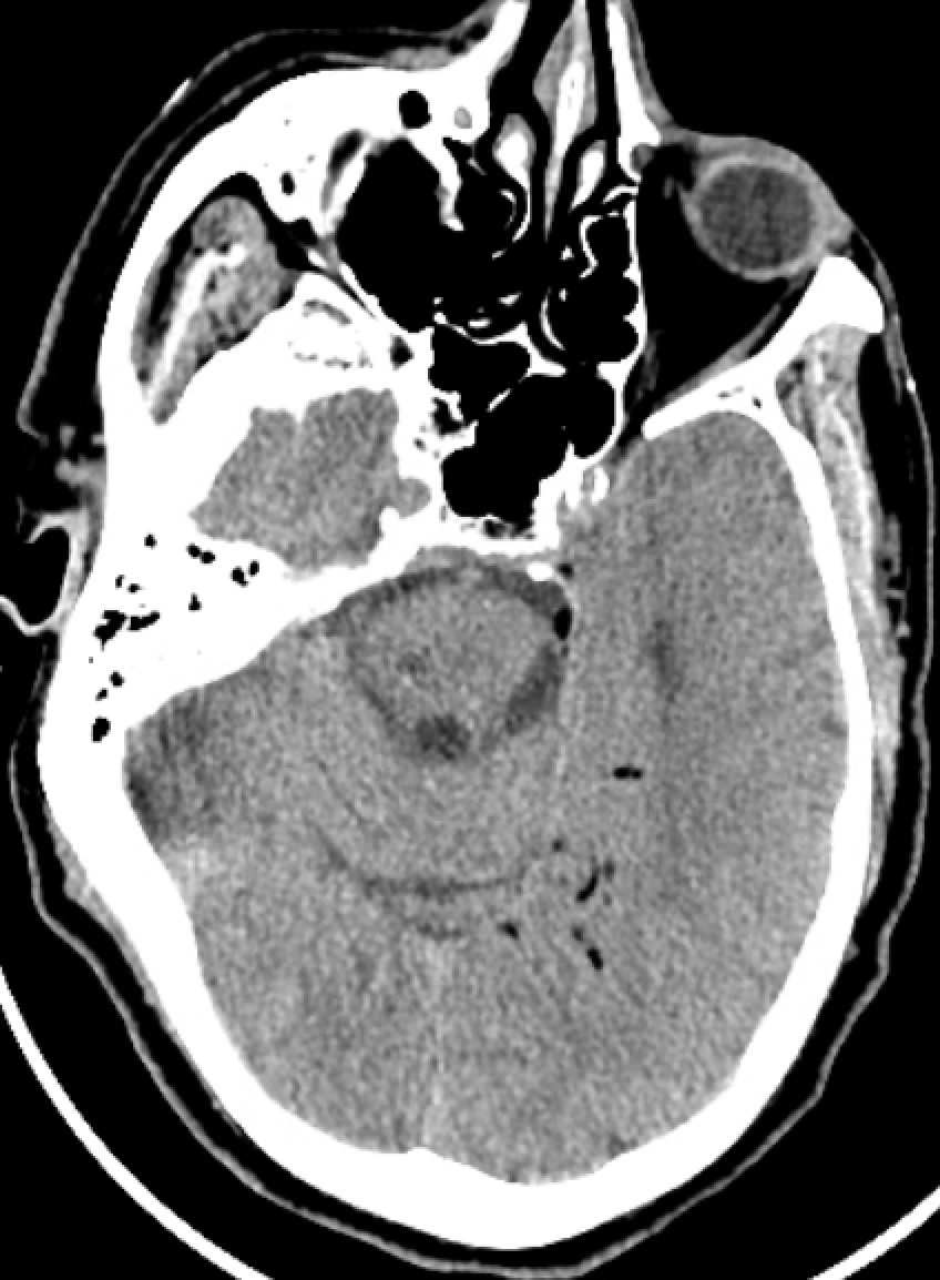
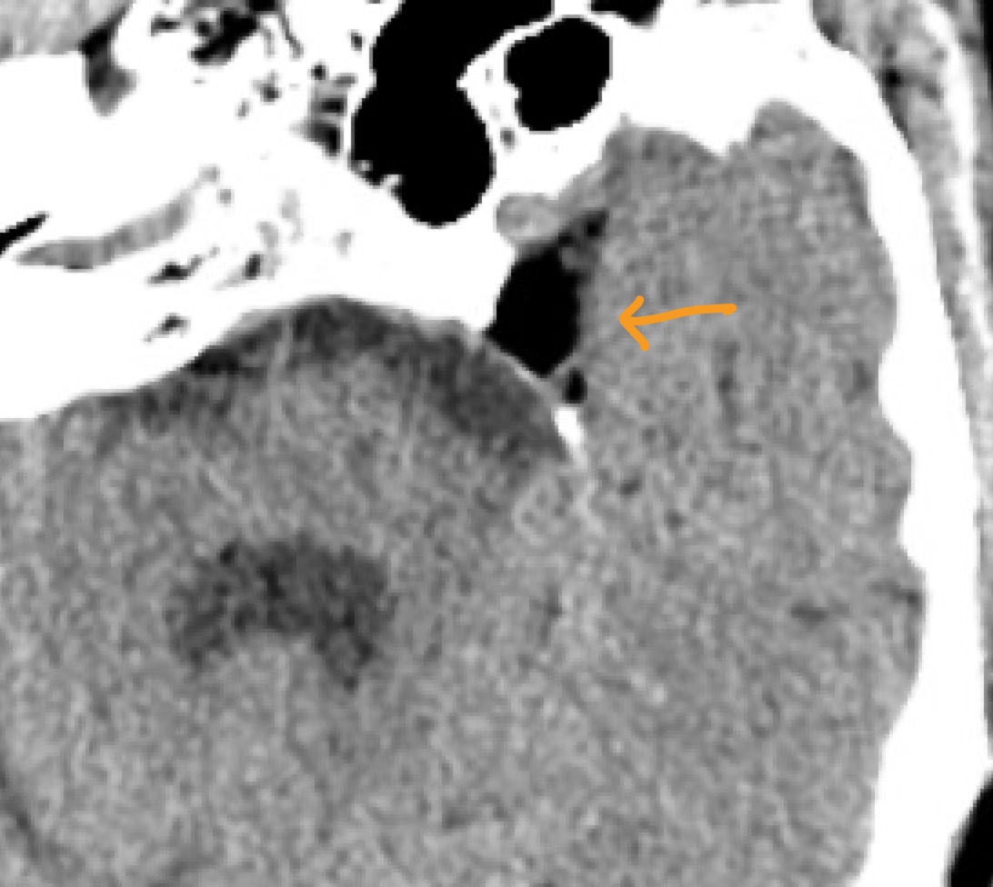
13/ Let’s now take a look at her MRI scan as well! Perhaps we'll see something interesting.
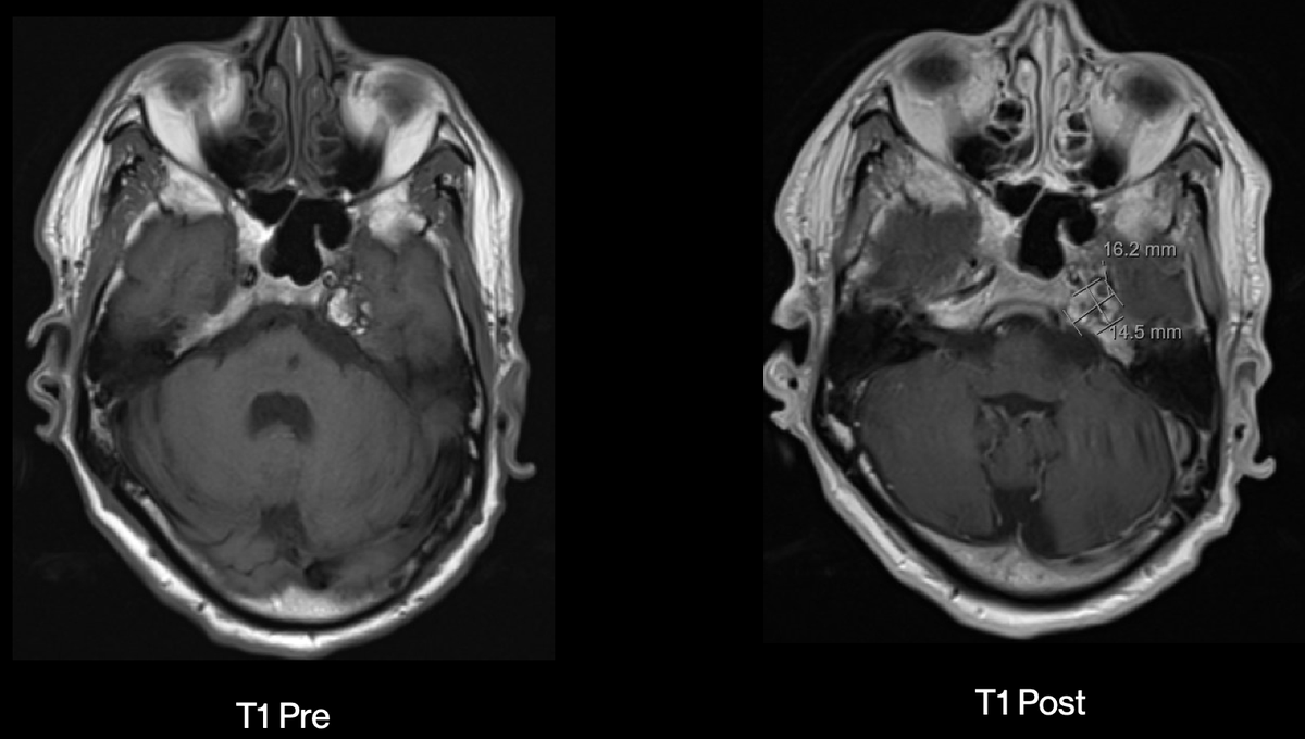
14/ Bingo, we have a ruptured dermoid cyst (red circle) that does not enhance post contrast!


15/ But a dermoid cyst!?
Let’s do a quick review ➡️ They account for <1% of all primary intracranial tumors and are often found incidentally (like ours). They occur as a developmental anomaly and usually occur at midline.
Let’s do a quick review ➡️ They account for <1% of all primary intracranial tumors and are often found incidentally (like ours). They occur as a developmental anomaly and usually occur at midline.
16/ They are usually benign tumors that lined by stratified squamous epithelium filled with mature mesodermal origin (hair follicles, sebaceous gland, etc).

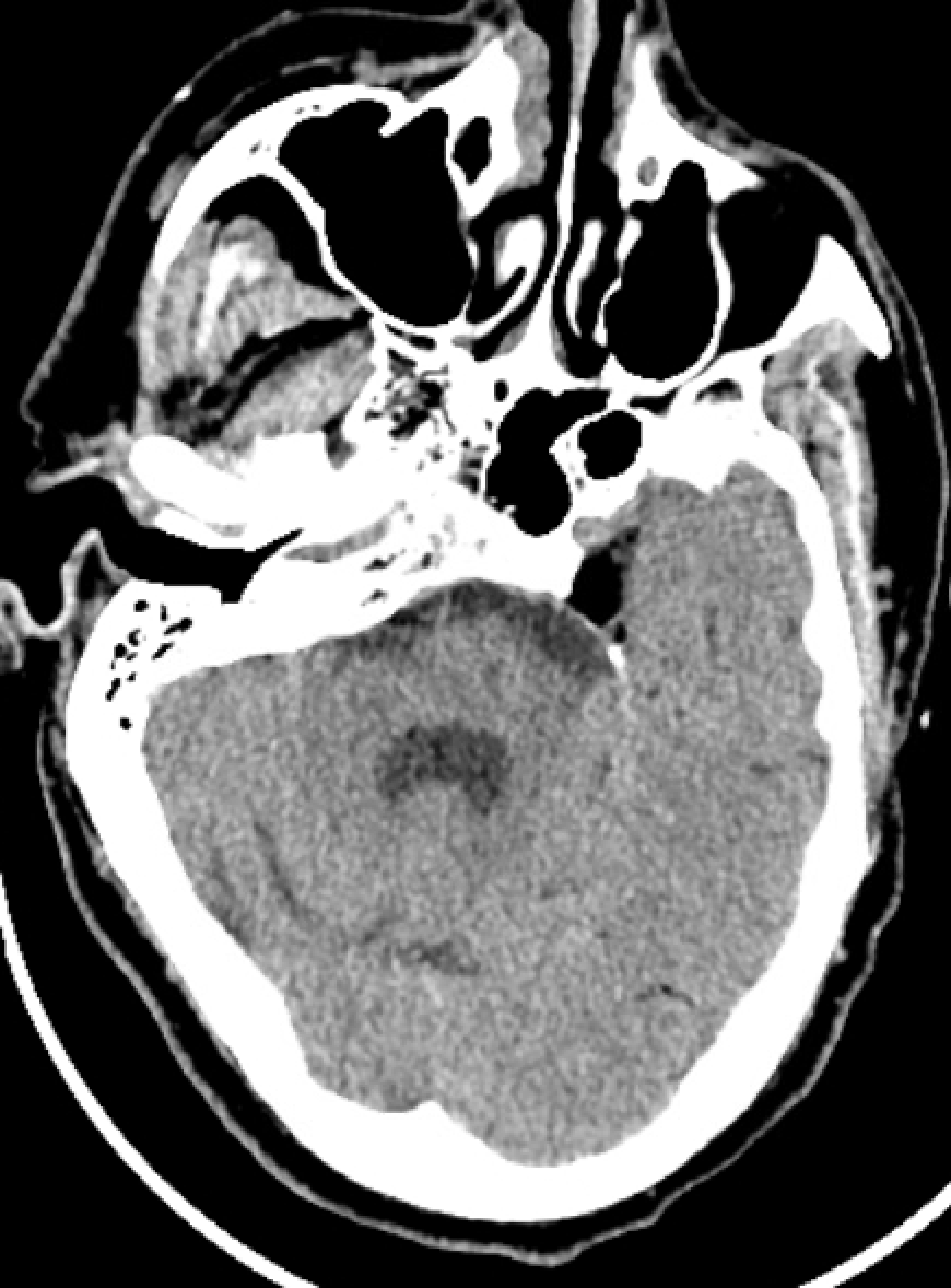
17/ Image wise, they appear as low attenuating masses on CT scan and characteristically are associated with fat attenuation material in the subarachnoid space upon rupture.
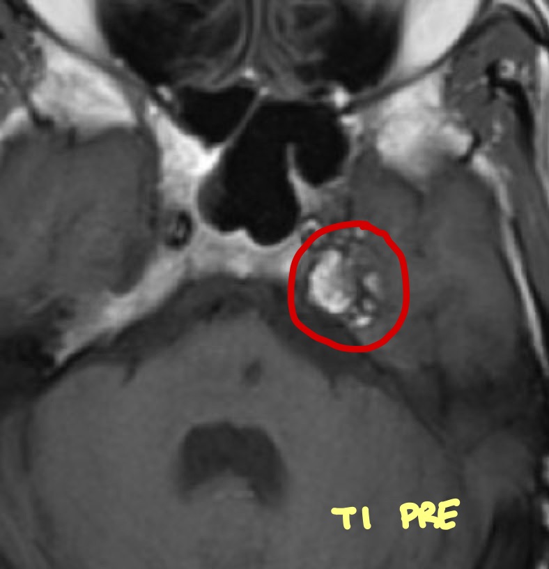
On MRI, they are hyperintense on T1 due to their cholesterol component and do not enhancement with GAD.
On MRI, they are hyperintense on T1 due to their cholesterol component and do not enhancement with GAD.

18/ For a vast majority, they can be left alone. However, when needed they can be surgically excised (which would also allow you to obtain a histological diagnosis).

19/ Here’s a great paper for a more detailed overview.
ncbi.nlm.nih.gov
While our patient didn't need any intervention, it was a good reminder that there's more to CT hypodensities other than just air.
ncbi.nlm.nih.gov
While our patient didn't need any intervention, it was a good reminder that there's more to CT hypodensities other than just air.
20/ Now the next time someone shows you a hypodensity on CT, make sure you sound like an expert radiologist and ask them “what’s the HU on it?" 😏

21/
🚨 And a special thanks as always to @caseyalbin 🚨
@sigman_md @mallyaa @EricLawson90 @maness_caroline @CajalButterfly @Capt_Ammonia @neuro_intensive @clhallmd @JackieKraft4 @feras_akbik @pouyeah @marcalainbabi @SubinMatthews @KP_MD2018 @rkchoi @WendeNGibbs @NMatch2022
🚨 And a special thanks as always to @caseyalbin 🚨
@sigman_md @mallyaa @EricLawson90 @maness_caroline @CajalButterfly @Capt_Ammonia @neuro_intensive @clhallmd @JackieKraft4 @feras_akbik @pouyeah @marcalainbabi @SubinMatthews @KP_MD2018 @rkchoi @WendeNGibbs @NMatch2022
Loading suggestions...